Imagine being able to spot the very first faulty wire in the brain — the loose connection that, years later, might unravel into Alzheimer's disease. A team of scientists in Illinois has just taken a meaningful step towards exactly that.
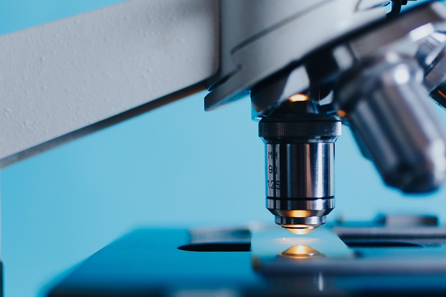
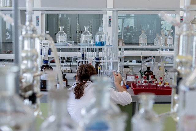
Researchers at the University of Illinois Urbana-Champaign have developed a technique that maps the connections between thousands of brain cells at once, with enough precision to see where individual neurons meet. The work, published in Nature Methods, could give doctors and drug developers a much sharper picture of what goes wrong in conditions such as Alzheimer's, Parkinson's and a range of psychiatric disorders — potentially long before a patient notices anything is amiss.
The trick involves giving each brain cell its own molecular label, a little like the barcode on a tin of soup. These labels are made from RNA, the same family of molecules that ferries genetic instructions around our cells. Specialised proteins carry the RNA barcodes from the body of the neuron out to the synapse — the tiny junction where one brain cell hands a signal to the next. When two neurons meet, their barcodes meet too. The researchers snip out those junctions, read the barcodes using high-speed genetic sequencing, and work out exactly which cells were talking to which.
The platform is called Connectome-seq, and its inventor is Boxuan Zhao, a professor of cell and developmental biology at Illinois.
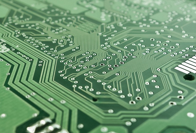
"When engineering a computer, you need to know the circuitry of the central processing unit. If you don't know how everything is wired together, you can't understand its function, optimise it or fix it when something breaks. We are approaching the brain the same way," Zhao said in a statement released by the university.
To explain how the barcodes work, he reaches for a homelier image. "Imagine a big bunch of balloons. The main body of each balloon has its unique barcode stickers all over it, and some move down to the end of the string. If two balloons are tied together at the end, the two barcodes meet at the junction," he said. "Then we snip out the knots and sequence the barcodes in each one. If the same knot has stickers from balloon A and balloon B, we know these two balloons are tied together."
A faster map of a famously complicated organ
Mapping the brain has, until now, been painstaking work. Scientists typically sliced tissue into wafer-thin sections, photographed each one under a microscope and pieced the pathways back together by hand. Newer sequencing methods could tag many neurons at once, but usually only showed roughly where a cell reached — not which partner it was actually wired to at the synapse.
Using Connectome-seq, Zhao's team mapped more than 1,000 neurons in a mouse brain circuit known as the pontocerebellar pathway, which links two regions involved in movement and coordination. The analysis turned up previously unknown links between cell types that nobody had realised were directly wired together in the adult brain, according to the University of Illinois.
Why patients might care
The hope is that, by comparing the wiring of healthy brains with those affected by disease, researchers will spot the earliest signs of trouble.
"With sequencing-based approaches, the time and cost are greatly reduced, which really makes it possible to see differences in different brains. We could see where connections change, where the most vulnerable parts of the brain are, perhaps before symptoms even appear," Zhao said. "For example, if we can catch where exactly the weak link is that kick-starts the whole catastrophic cascade in Alzheimer's disease, can we specifically strengthen those connections to where the disease slows or does not progress?"
For now, Connectome-seq has been demonstrated only in mice, and Zhao's team is still working to scale it up to map a whole mouse brain. Human applications are some way off. But for a field that has spent decades squinting at the brain one slice at a time, a method that can read thousands of connections in parallel is, quietly, a big deal.
The work was supported by the Wu Tsai Neurosciences Institute at Stanford University, the Elsa U. Pardee Foundation and the Edward Mallinckrodt Jr. Foundation.